Chiropractic care for the cervical spine as a treatment for plagiocephaly: a prospective cohort study
By Nicola Ann Douglas, MChiro, MSc1, Maria Browning, BSc, DC, MSc (Chiro Paeds), Cert Med1,
Joyce Miller, BS, DC, PhD2
1. Private practice, United Kingdom
2. Corresponding author: Joyce Miller, clinic tutor, Anglo-European College of Chiropractic (AECC), Bournemouth, United Kingdom
Email: JMiller@aecc.ac.uk
Abstract
Background: Plagiocephaly is a condition that affects the shape of the skull in infants. The research has suggested that there is a growing association between plagiocephaly and developmental delay later in infancy extending into childhood. Plagiocephaly is an increasingly common condition in society which often presents to a chiropractic practice. There have been no studies prospectively investigating the outcome of chiropractic care on a group of infants with plagiocephaly. Objectives: To observe any change in head deformation measurements in a single cohort group of infants aged 0-12 months old presenting to a chiropractic clinic and receiving chiropractic care over a course of six weeks. Setting: This single cohort observational study took place at a chiropractic teaching clinic between February and July 2015, on the south coast of England. Methods: Infants presenting to the chiropractic clinic with the complaint of plagiocephaly were measured from the external occipital protuberance (EOP) to the anterior ear both right and left sides during their routine course of treatment. These measurements were re-examined at the end of their course of care or at six weeks after presentation, which ever occurred first. Results: A total of 64 infants were included. The mean change in the plagiocephaly measurement was a reduction of 1.13cm ± 0.89cm, p= 0.00, 95%CI (-1.36 to -0.92cm). Overall, 20 out of the 64 participants showed a full resolution of plagiocephaly, with a final measurement below 0.4cm difference in occipital measurement side to side, which is considered normal. No adverse events were reported for any of the infants. Conclusions: Overall, there was both a statistically and clinically significant reduction in plagiocephaly measurement for this cohort of infants after a course of chiropractic care. As this was an observational study, this cannot be interpreted as cause and effect. However, these results encourage further research, particularly a RCT to investigate the effect of chiropractic care on plagiocephaly in infants.
Introduction
Plagiocephaly is a term used to describe an asymmetry in the shape of the skull. Non-synostotic plagiocephaly describes a flattening of the skull that evolves into two types of shape deformities (Figure 1). Firstly, the posterior-lateral occiput can appear flattened, termed plagiocephaly and secondly a uniform flattening across the back of the occiput is termed brachycephaly.1

Figure 1: A comparison of the two different head asymmetries in positional plagiocephaly, amended with permission from Saich and Laker 2014.37
The cause of plagiocephaly is exposure of the cranium to external compressive forces while it is still developing. The flatness is usually first recognised by parents when the infant is 2-3 months old.2 Since the ‘Back to Sleep Program’ in 1992 there has been a significant rise in the prevalence of plagiocephaly. However the actual prevalence of plagiocephaly is uncertain due to the range in estimates. Estimates vary from 16-21% at 6-7 weeks old and 20% at 4 months old3 to 47% of infants aged 7-12 weeks old out of cohorts of healthy full term infants.4 The real issue is not only the rise in incidence but also long-term risk factors. The UK National Health Service (NHS) states plagiocephaly is “cosmetic only.”5 This statement contradicts the research suggesting that plagiocephaly is associated with learning disabilities, dysfunctional auditory processing and developmental delay in the gross motor and cognitive domains.6,7,8,9,10
There is further conflict of opinion as to whether plagiocephaly naturally resolves as the head develops or whether interventions may be helpful to change the long-term course of the condition.10,11 There have been several studies comparing the effects of cranial orthosis against physical therapy or active counter positioning therapy on plagiocephaly in infants. However, there is no evidence of benefit for cranial orthoses or helmets in the treatment of plagiocephaly.1
The research suggests that physical therapy or active counter positioning may be effective when the infants are young with mild to moderate plagiocephaly.12 One randomized controlled trial (RCT) found active counter positioning advice to parents/guardians had a positive and significant effect on plagiocephaly in infants compared to no instruction.13 It is inconclusive whether physical or manual therapy may be more important than what is currently understood for the appropriate management of plagiocephaly which is no clinical management. The purpose of this investigation was to assess whether there is a role for chiropractic manual therapy in the routine treatment of plagiocephaly in a clinical population.
Background
There is little research investigating the effect of chiropractic care, a form of manual therapy, on plagiocephaly. There has been one retrospective study14 and several case studies.15,16,17,18,19 Also there has been one pilot single cohort study investigating the effect of osteopathy, another form of manual therapy on plagiocephaly in infants.20 All of these studies showed improvement in plagiocephaly in infants.
Chiropractors commonly manage infants with plagiocephaly and treat the cervical spine to achieve full range and freedom of motion so that the infant is not “stuck” in one position. If the head can be freely moved, then the pressure on the cranium will be equally distributed and the skull will, ideally, become symmetrical.
This is an appropriate method for resolution as research has shown there is a relationship between cervical range of motion and plagiocephaly.7,21,22 Decreased cervical motion was found to be associated with a larger cephalic index (calculated by cranial width divided by cranial length, multiplied by 100) and brachycephaly.21 It seems intuitive, but research has also found that infants with head positional preference are four times more likely to have plagiocephaly than infants without positional preference.22 Their study found that the condition is at increased risk when infants do not vary their head position and leads to one question: is this because they are unable to vary their head position? This tendency has also led to referring to plagiocephaly as positional head deformation (PHD). We found this easier to say and understand by the parents as well as more descriptive of the condition and we use that term clinically.
The link of head deformation to suboptimal cervical spine rotation has been corroborated with research that has shown that physical therapy improved cervical range of motion in infants with plagiocephaly and this significantly improved cranial symmetry compared to a control group receiving counter positioning advice only.23 Based on those findings it was recommended to general practitioners to consider manual therapy for the early management of infants with plagiocephaly.24 Despite the fact that chiropractors are known for manual therapy and are the most common choice for families choosing CAM care for their infant,25 there is still a great need for further research investigating the outcomes of chiropractic care with positional head deformation prospectively in a clinical setting.
Method
This study was a prospective cohort study conducted at the Anglo-European College of Chiropractic (AECC) clinic, Bournemouth, UK. The AECC ethics subcommittee granted ethical approval in January 2015.
A literature search was conducted using the online databases of Cochrane Library, Index to Chiropractic Literature, Pubmed and Science Direct. Key words were ‘deformational plagiocephaly,’ ‘positional plagiocephaly’ and combined individually and together with ‘chiropractic,’ ‘infant’ and ‘manual therapy.’ Publications of RCTs, systematic reviews, case studies, cohort retrospective and prospective studies were included. Publications involving craniosynostosis or surgical cases were excluded as well as publications before 2001 to allow for the most current literature. Thirty-one articles were found and obtained through the online and library resources at Bournemouth University and were reviewed. A cross-reference search was also conducted for any relevant but unfound studies at the end of each paper. Several studies were relevant but were all included in the databases search. A hand search for relevant but unpublished studies and this was also conducted at Bournemouth University; one thesis was found.
Population
The sample group of participants were infants aged between 0-12 months old, diagnosed with plagiocephaly whose parents presented their child to the AECC clinic for care and gave their consent for the infant to receive chiropractic treatment. Participant data was excluded from this observational study if the infant was diagnosed with craniosynostosis, or undergoing any other form of medical treatment or therapy.
Procedure
The routine chiropractic examination at the AECC clinic involves a musculoskeletal assessment recording head shape as well as other key measurements such as vital signs documented in each infant’s file. Positional Head Deformation (PHD) was determined by two cranial measurements taken from the external occipital protuberance (EOP) to the anterior ear for both left and right side (Image 1). A difference of more than 0.4cm between the two sides was recorded as plagiocephaly or PHD present.21 These measurements were taken using the prototype occipital measurement device (Image 2) that has shown to have a high inter-examiner reliability.26 Following the standard AECC clinic’s protocol for treatment of infants and children, these measurements were taken at the 1st, 4th and 7th treatment visit, and/or at the date the child was released from care or monitored over a maximum time frame of six weeks. Six weeks was chosen because reduction in that time frame would be considered well ahead of the natural history of the disorder.
 |
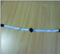 |
Image 1: A photograph of measuring the infant’s EOP
to anterior ear using the prototype occipital measuring device. |
Image 2: The prototype occipital measuring device. |
Following a routine course of chiropractic care for infants with plagiocephaly, the infants’ data were gathered from each of their files. Confidentiality for each participant remained intact throughout the study which was purely observational as the condition is known to have long-term negative sequelae. There was no control or comparison group that did not receive care, as this could be considered unethical in the light of the negative long-term prospects associated with the condition.6,7,8,9,10
Results
A total of 64 participants met the inclusion criteria. The clinical characteristics of the participants are shown in Table 1. There were more males than females, 62.5% (n=40) and 37.5% (n=24) respectively. The age range of infants presenting to the clinic varied from 2 to 44 weeks old, average age was 11.5 ±6.76 weeks old, 95%CI (9.84 to 13.2weeks). 63 of the 64 participants were aged under 6 months with the exception of one participant aged 44 weeks (11 months) old.

This study found that there were differences in the record keeping so there were different numbers who answered specific questions. The number (n) is shown in the tables, along with percentage. On average, most infants were full-term with 56% assisted births and 44% natural vaginal deliveries. All of the infants had a limitation in cervical spine range of motion, with 46% having preference to rotate to the right, 40% to the left and 4% where parents did not know the direction of their child’s preference.
The average difference in side-to-side EOP to anterior ear measurement for this group of infants before chiropractic treatment was 1.71± 0.84cm, 95%CI (1.50 to 1.91cm). At the end of the course of chiropractic care these measurements reduced to 0.67 ± 0.65cm, 95%CI (0.50 to 0.83cm), (Figure 2). The mean change in the occipital measurement was a reduction of 1.13cm ± 0.89cm, p=0.000, 95%CI (-1.36 to -0.92cm), on average a 33% decrease in difference side to side. Overall, 20 out of the 64 participants showed a full resolution of head deformation with a measurement difference below 0.4cm.

Figure 2: Plagiocephaly measurements before and after chiropractic treatment.
All participants (n=64) had full range of cervical motion restored following the course of chiropractic care. On average, the course of chiropractic care consisted of five treatments over a period of six weeks. The infant’s head circumferences changed throughout the course of chiropractic care as one would expect with growth. The average head circumference for 21 participants in this study’s sample of infants at the start of the study was 40.6cm ± 2.10, 95%CI (39.7 to 41.5cm) that increased to 42.2cm ± 2.56, 95%CI (41.1 to 43.3cm) at the end of the study. Overall there was a mean increase of 1.61cm ± 1.68, 95%CI (0.89 to 2.33cm).
Discussion
This study was designed to observe infants presented for care for positional head deformation by their parents over a course of chiropractic treatment lasting up to six weeks.
This study’s population was representative of infants with PHD. For example there were more boys than girls. Plagiocephaly is more prevalent in males.2 However, another recent study found plagiocephaly was present in an equal number of males and females in a healthy cohort of neonates21 and this is likely due to the general increased prevalence of plagiocephaly.4 It must be noted that the Aarivala et.al. 2014 study included only neonates and perhaps it is that both genders have equal amounts of head deformation as newborns but that it is more likely to persist in males. This concept has never been studied.
Also, the majority of the participants were under six months (98% n=63) with half between 2-4 months old; other studies report that plagiocephaly is most prevalent between 2-4 months old.1,27,28 One explanation is due to the acceleration in the infant’s head development at this age. Any excessive compression to one aspect of the skull will alter the shape and appear as flattened in comparison to the rest of the skull’s normal development.21
Another example that this study’s population was representative of the general PHD population, was a quarter of the subjects had been born prematurely, which is higher than the average incidence in developed countries of 5-9%.29 Other research suggests that prematurity is over-represented in children with misshapen heads.28,30
The same is true for over-representation of assisted births which accounted for over half of the births in this cohort. The average in the UK population is 39%.31 Plagiocephaly is associated with assisted births.21
Almost ¾ of this sample were first borns and ¼ were twins, far outnumbering averages for routine births in the UK population (equal ratios between first born, second born and third or more born in the UK32 and 1.52% of all births in the UK were multiple births of twins in 2012).33
Cervical mobility restrictions were reported in all of the infants in this sample. Research has suggested when the infant’s neck has limited movement, the infant will have a preference for a particular side to turn as they are unable to turn equally to both right and left.7,21 These findings also support research that cervical restriction and plagiocephaly are linked with neurological disruption and developmental delay due to factors associated with inactivity and variable tone.7,9,34 It is unlikely that the association with developmental delay is due to the head deformation, but more likely to be related to the poor movement patterns associated with it. This highlights the need for manual therapy to restore normal range of motion and beneficial movement.
The infants’ plagiocephaly measurement significantly reduced over the course of chiropractic care. Out of the sample of 64 participants, 20 showed a complete resolution of head deformation, a value of 0.4cm or less difference between left and right side which is widely recorded as a normal, almost imperceptible difference.2 The remaining infants showed a reduction in the plagiocephaly measurement with a mean reduction of 1.13cm ± 0.89cm, p=0.000, 95%CI (-1.36 to -0.92cm). That improvement was both statistically and clinically significant and shows a percentage decrease of one third and the improvement would be expected to continue with head growth, with the impediment to cervical range of motion removed.
Chiropractic treatment involved pediatric manipulative therapy to the spine and extremities through the form of press and hold methods aimed to mobilize any joints involved, a low force treatment of 2-8 newtons depending on the age and condition of the child. The chiropractor also gave advice to parents on supervised prone playing (tummy time) for their infant, as well as home measures for active counter positioning to encourage the child to look to all directions. Although there are no clear updated guidelines on prone play (tummy time) for infants, the AAP (1998)35 did suggest that it was important to start the first week with awake-time repositioning as the supine sleep program had resulted in an epidemic of head shape problems. There were no negative side effects or adverse events reported during the course of chiropractic care.
There are limitations to this type of study. Not only is there not a control group for comparison but this is a time period when infant’s heads undergo accelerated growth. In fact, this factor most likely contributes to the rapid improvement in head shape. The average head circumference is 35 cm in full term neonates increasing to 45 cm by 1 year old and 55cm in a grown adult.36 Therefore, there is rapid head growth within the first year of the infant’s life. Head circumference increases, on average, by 2cm per month for the first 3 months of life, 1 cm per month for the second 3 months of life and 0.5cm per month from 6-12 months.36 However, there is little evidence that head shapes change along with growth unless the infant is able to perform full cervical range of motion and compress both sides of the occiput evenly. It does seem as though a key to improvement is to facilitate full cervical range of motion, which is what the chiropractic care was designed to do.
That said, the plagiocephaly measurement could have reduced naturally given significantly more time. It has been discussed in previous studies that infant’s head shapes return to their normal shape by 3-5 years of age.1,11,13 Other research has found contrary findings and suggested plagiocephaly remains further into childhood.10 Therefore the reduction in plagiocephaly seen amongst this sample of infants could have been down to their natural head development. However the research has suggested this process occurs later in infancy and early childhood instead of during early infancy. It can be said that the changes in this study have occurred ahead of the natural history of the disorder.
Further, all infants received the full spectrum of chiropractic care which involved manual therapy, along with advice for counter positioning and tummy time. Therefore, the positive effects found after chiropractic care could have been the effect of counter positioning and tummy time measures given by the parents instead of the application of chiropractic manual therapy. Counter positioning measures have been found to have positive outcomes of a reduction in plagiocephaly measurements in infants when compared to cranial orthosis12 and as a measure on its own.13 However, research has also shown that compared to physical therapy, greater effects were seen in the physical therapy group23 in two groups of infants with plagiocephaly. There have been no studies prospectively investigating the effect of chiropractic care on plagiocephaly in infants compared to parent measures. In future studies it would be interesting to compare the two therapeutic measures and have two groups of infants with plagiocephaly, one group receiving chiropractic care and one counter positioning techniques. However, ethical consideration is required with this type of study and since there is some evidence that both work, it may not be ethical to withhold any type of effective treatment, considering the long term negative associations with unresolved plagiocephaly.
Another limitation to this study was the prototype measurement device used to determine the plagicoephaly value. This has been shown to have high inter examiner reliability26 but it has not been validated. There is also the limitation of human error and the clinicians using this device may have not correctly read the infants’ EOP to anterior ear measurement. This prototype measurement device was used as it was easy and inexpensive to implement in a routine clinical setting compared to other anthropometric or digital measuring devices. However, by choosing this device it does have its’ limitations such as the measurement from EOP to anterior ear difference between left and right side would only show one form of plagiocephaly, positional plagiocephaly instead of brachycephaly as well. Also the prototype measurement device is not a standardized device for measuring plagiocephaly, therefore this study is more difficult to compare to other studies using different devices. However, parental perception of improvement is at this time the gold standard for outcomes, and this device compared favourably to simple observation by parents and clinicians as well as supplying some level of objectivity.
Conclusion
This study showed a significant reduction in head deformity in infants during a course of six weeks of chiropractic care. Although this cannot be interpreted as cause and effect because the research was observational in a routine clinic setting rather than a randomized controlled trial, the improvements in head shape occurred earlier than the natural course of the condition. Further research is strongly encouraged to investigate the effects of chiropractic management for this condition.
References
1. van Wijk RM, van Vlimmeren LA, Groothuis-Oudshoom CG, Van der Ploeg CP, ljzerman MJ, Boere-boonekamp MM. Helmet therapy in infants with positional skull deformation: randomized controlled trial [Internet]. BMJ 2014 May 1; [cited 2015 Oct 9]. Available from: http://www.bmj.com/content/348/bmj.g2741.long.
2. Hutchinson BL, Hutchinson LAD, Thompson JMD, Mitchell EA. Plagiocephaly and brachycephaly in the first two years of life: a prospective cohort study. Pediatrics 2004; 114(4):970-80.
3. Bialocerkowski AE, Vladusic SL, Wei C. Prevalence, risk factors and natural history of positional plagiocephaly: a systematic review. Dev Med Child Neurol 2008; 50(8):577-86.
4. Mawji A, Vollman AR, Hatfield J, McNeil DA, Sauvé R. The Incidence of positional plagiocephaly: a cohort study. Pediatrics 2013; 132(2);298-304.
5. National Health Service. Plagiocephaly and brachycephaly (flat head syndrome) [Internet]. London: National Health Service; 2014 April 29 [cited 2015 13 Oct]. Available from: www.nhs.uk/conditions/plagiocephaly/Pages/Introduction.aspx.
6. Collett BR, Breiger D, King D, Cunningham ML, Speltz ML. Neurodevelopmental implications of “deformational” plagiocephaly. J Dev Behav Pediatr 2005; 26(5):379-89.
7. Hutchinson BL, Stewart AW, Mitchell EA. Characteristics, head shape measurements and developmental delay in 287 consecutive infants attending a plagiocephaly clinic. Acta Paediatr 2009; 98(9):1494-9.
8. Speltz ML, Collett BR, Scott-Miller M, Starr JR, Heike C, Wolfram-Aduan AM, King D, Cunningham ML. Case-control study of neurodevelopment in deformational plagiocephaly. Pediatrics 2010; 125(3):537-542.
9. Collett BR, Starr JR, Kartin D, Heike CL, Berg J, Cunningham ML, Speltz ML. Development in toddlers with and without deformational plagiocephaly. Arch Pediatr Adolesc Med 2011; 165(7):653-8.
10. Collett BR, Gray KE, Starr JR, Heike CL, Cunningham ML, Speltz ML. Development at age 36months in children with deformational plagiocephaly. Pediatrics 2013; 131(1):109-15.
11. Hutchinson BL, Stewart AW, Mitchell EA. Deformational plagiocephaly: a follow-up of head shape, parental concern and neurodevelopment at ages 3 and 4 years. Arch Dis Child 2011; 96(1):85-90.
12. Kack Flannery AB, Looman WS, Kemper K. Evidence-based care of the child with deformational plagiocephaly, Part II: Management. J Pediatr Health Care 2012; 26(5):320-31.
13. Hutchinson BL, Stewart AW, De Chalain TB, Mitchell EA 2010. A randomized controlled trial of positioning treatments in infants with positional head shape deformities. Acta Paediatr 2010; 99(10):1556-60.
14. Davies NJ. Chiropractic management of deformational plagiocephaly in infants: an alternative to device-dependent therapy. Chiropractic Journal of Australia 2002; 32(2):52-5.
15. Leighton JM. Non-synostotic deformational plagiocephaly: an evidence-based case report. Clinical Chiropractic 2008; 11(4):211-8.
16. Fairest C. Resolution of birth-related cranial asymmetry following chiropractic care: a case report. Journal of Clinical Chiropractic Pediatrics 2013; 14(1):1093-6.
17. Generaux CD, Alcantara JD. Resolution of birth induced facial asymmetry following chiropractic care: a case report. J Pediatr Matern & Fam Health 2011 (Winter):35-9.
18. Hash JL. Deformational plagiocephaly and chiropractic care: a narrative review and case report. Journal of Clinical Chiropractic Pediatrics 2014; 4(2):1132-8.
19. Humphris TJ, Askin ASMM, Glucina-Russell TT. Resolution of non-synostotic plagiocephaly flowing chiropractic care: a case report. Journal of Clinical Chiropractic Pediatrics 2014; 14(3):1176-8.
20. Lessard S, Gagnon I, Trottier N. Exploring the impact of osteopathic treatment on cranial asymmetries associated with nonsynostotic plagiocephaly in infants. Complementary Therapies in Clinical Practice 2011; 17 (4):193-198.
21. Aarnivala HEI, Marita Valkama A, Pirttniemi PM. Cranial shape, size and cervical motion in normal newborns. Early Hum Dev 2014; 90(8):425-30.
22. Mawji A, Vollman AR, Fung T, Hatfield J, McNeil DA, Sauvé R. Risk factors for positional plagiocephaly and appropriate time frames for prevention messaging. Paediatr Child Health 2014; 19(8):423-7.
23. van Vlimmeren LA, van der Graaf Y, Boere-Boonekamp MM, L’Hoir MP, Helders PJ, Engelbert RH. Effect of pediatric physical therapy on deformational plagiocephaly in children with positional preference, a randomized controlled trial. Arch Pediatr Adoles Med 2008; 162(8):712-8.
24. Vargish L, Mendoza MD, Ewigman B. Use of physical therapy to head off this deformity in infants. J Fam Pract 2009; 58(8):2-3.
25. Kemper KJ, Vohra S, Walls R, task force on complementary and alternative medicine, provisional section on complementary and integrative medicine. American Academy of Pediatrics. The use of complementary and alternative medicine in pediatrics. Pediatrics 2008;122(6):1374-86.
26. Breen JJ, Browning M. Interexaminer reliability of measurements for positional head deformity in infants in a clinical setting. Thesis (MSc) 2013; Bournemouth University.
27. Boere-Boonekamp MM, Linden-Kuiper LT. Positional preference: prevalence in infants and follow up at 2 years. Pediatrics 2001; 107(2):339-43.
28. Bialocerkowski AE, Vladusic SL, Wei C. Prevalence, risk factors and natural history of positional plagiocephaly: a systematic review. Dev Med Child Neurol 2008; 50(8):577-86.
29. Beck S, Wojdyla D, Say L, Betran AP, Merialdi M, Requejo JH, Rubens C, Menon R, Van Look PFA. The worldwide incidence of preterm birth: a systematic review of maternal mortality and morbidity. Bull World Health Organ 2010; 88(1):31-8.
30. Hutchinson BL, Thompson JMD, Mitchell EA. Determinants of nonsynostotic plagiocephaly: a case-controlled study. Pediatrics 2003; 112 (4):316-322.
31. Health and social care information centre (HSCIC). NHS maternity statistics 2013-14 [Internet]. Leeds: Health and social care information centre; 2015 Jan 28 [cited 2015 May 25]. Available from:
http://www.hscic.gov.uk/catalogue/PUB16725.
32. Department of Health (DOH). Birth ratios in England and Wales: a report on gender ratios at birth in England and Wales [Internet]. London: Department of Health; 2014 Oct 5 [cited 2015 Oct 12]. Available from: https://www.gov.uk/government/uploads/system/uploads/attachment_data/file/313559/Sex_Selection_in_England_and_Wales_analysis_April_2014_NM_comments.pdf.
33. Office for National Statistics (ONS). Birth registrations, place of birth and multiple births in England and Wales: 2013 [Internet]. London: Office for National Statistics; 2014 Nov [cited 2015 Oct 12]. Available from: http://www.ons.gov.uk/ons/index.html.
34. Fowler EA, Becker DB, Pilgram TK. Neurologic findings in infants with deformational plagiocephaly. J Child Neurol 2008; 23(7):742-7.
35. American Academy of Pediatrics Task Force on Infant Positioning and SIDS Update. Pediatrics 1998; 98(6):1216-1218.
36. Bronfin DR. Misshapen heads in babies: position or pathology. Ochsner J 2001; 3(4):191-9.
37. Saich P, Laker K. Raising awareness of plagiocephaly and brachycephaly: diagnosis [Internet]. Cambridge: Headstart4babies; 2014 [cited 2015 Oct 14]. Available from: http://www.headstart4babies.org.
|
